
Fig. 8.
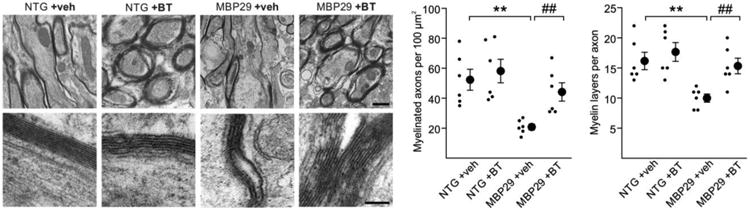
Structural restoration of myelin by benztropine in mice overexpressing α-synuclein in oligodendrocytes (MBP29). Representative electron micrographs of the corpus callosum at low (upper panel, scale bar: 1μm) and high (lower panel, scale bar: 0.2μm) magnification depict myelin sheaths of non-transgenic (NTG) and MBP29 mice treated with either vehicle (veh) or benztropine (BT). In vehicle-treated MBP29 mice only, myelin appeared strongly reduced and disorganized. Quantification of myelinated axons and myelin layers per axon revealed a significant myelin deficit in vehicle-treated MBP29 mice, which was restored by benztropine treatment to control level. Data are shown as mean ± standard error of mean. ANOVA (n = 6): **p < 0.01. T-test: ##p < 0.01.