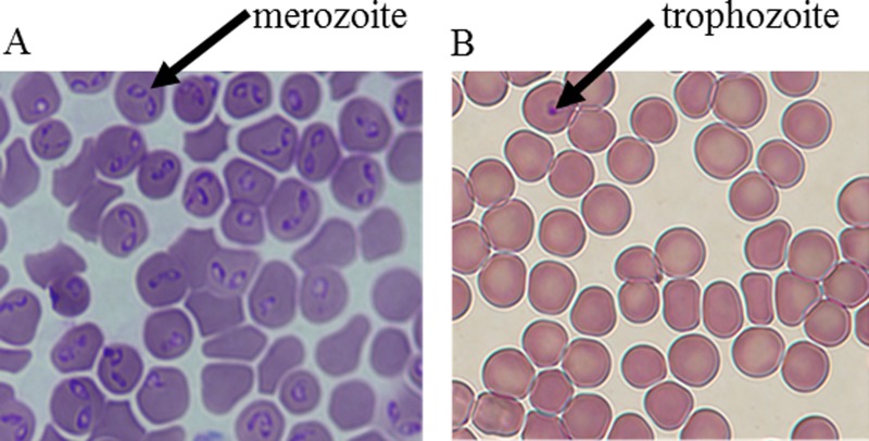
FIG. 5.
Images obtained at 100× magnification using post-separation diff-quik-stain kit showing Babesia-infected and healthy erythrocytes. Only the parasites were stained. Here, the cells were stained after they had been separated. (a) Microdevice outlet port 1 (rich in Babesia-infected erythrocytes). (b) Microdevice outlet port 2 (lean in Babesia-infected erythrocytes). The arrows show the parasite (nucleus-like structure) residing in the RBCs. The pair-shaped parasites are the merozoites while the round-shaped parasites are the trophozoites. Any RBC that contains the parasite is considered infected. The images were obtained when the inlet parasitemia was ∼5%.

