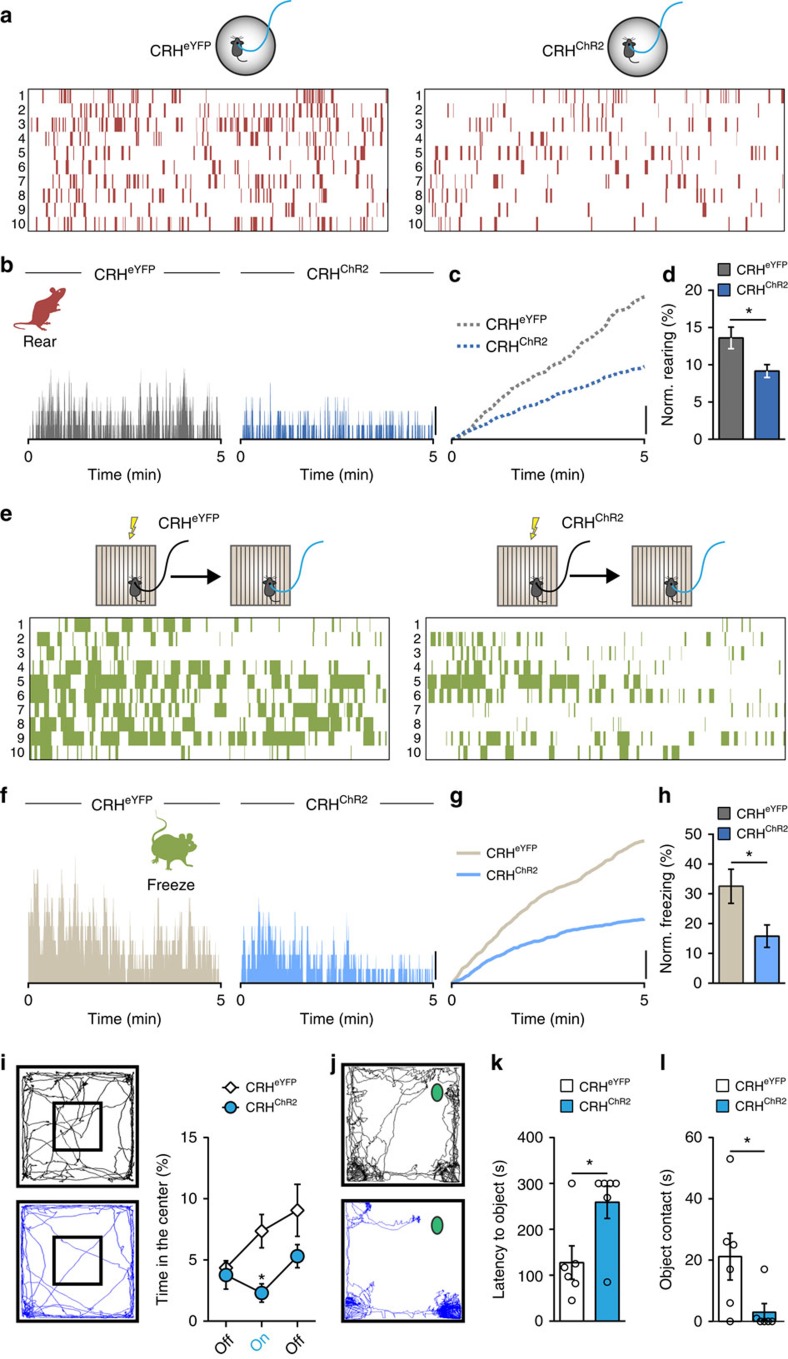
Figure 7. Photostimulation of PVN CRHChR2 neurons overrides contextual cues.
(a–d) Optical stimulation of PVN CRH neurons attenuates rearing in novel environment (Novel). (a) Each row represents an individual animal. (b) Histograms showing percentage of animals rearing. (c) Cumulative graphs demonstrate the relative extent of rearing. (d) Rearing time as a fraction of all behaviours after exclusion of time spent grooming (CRHeYFP: 13.6±1.4%, n=10; versus CRHChR2: 9.2±0.9%, n=10; P=0.0165; t-test). (e–h) Optical stimulation of PVN CRH neurons disrupts freezing in FS. (e) Each row represents an individual animal. (f) Histograms show percentage of animals freezing. (g) Cumulative graphs demonstrate the relative extent of freezing. (h) Quantification of fractional freezing time if time spent grooming is excluded from the analysis (CRHeYFP: 32.5±5.7%, n=10; versus CRHChR2: 15.8±3.8%, n=10; P=0.0251; t-test). (i) Assessment of locomotion in an open field test. Representative locomotor trajectory plots during optical stimulation in CRHeYFP (black) and CRHChR2 (blue) mice. Accompanying graph shows CRHChR2 mice spend significantly less time spent in the centre zone during photostimulation (CRHeYFP: before: 3.9±0.5%, during: 6.1±0.5%. after: 6.6±0.9, n=16; versus CRHChR2: before: 4.3±1.0%, during: 3.3±0.8%, after: 5.3±0.8%, n=14; CRHeYFP during versus CRHChR2 during, P=0.0291; repeated-measures two-way ANOVA). (j) Representative locomotor trajectory plots during optical stimulation in CRHeYFP (black) and CRHChR2 (blue) mice in a novel object (green shape) test. Optical stimulation reduces exploration of a novel object as measured by the latency to touch (k, CRHeYFP: 127.7±36.4 s, n=6; versus CRHChR2: 259.0±35.2 s, n=6; P=0.0267; t-test) and the time spent in close proximity (l, CRHeYFP: 21.2±7.6 s, n=6; versus CRHChR2: 3.0±2.8 s, n=6; P=0.0494; t-test). Scale bars: (b,c,f,g), 20%; *P<0.05; Error bars±s.e.m.

