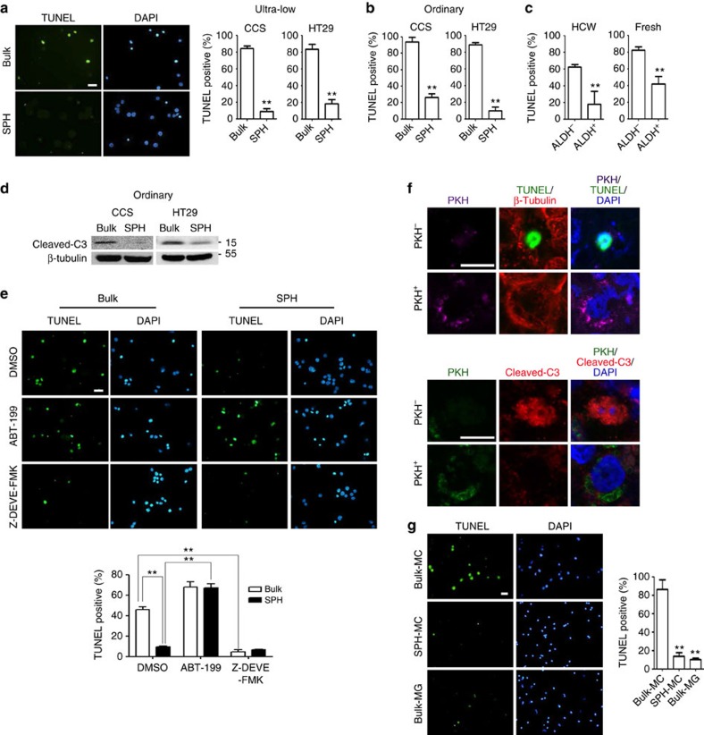
Figure 2. Spheroid culture-enriched TICs show better suspension-survival ability.
(a) Left, representative pictures of TUNEL staining for HT29. Right, quantification of TUNEL-positive cells. (b,d) Bulk cancer cells (Bulk) and spheroid culture-enriched TICs (SPH) were cultured under spheroid condition for (b) 24 h and (d) 10 h, followed by (b) TUNEL assay and (d) western blot analysis, respectively. (c) ALDH− and ALDH+cells isolated from HCW primary liver metastasis colorectal cancer cells were cultured under spheroid condition for 24 h, followed by TUNEL assay. (e) HT29 bulk cancer cells and spheroid culture-enriched TICs were cultured under spheroid condition in the presence of Bcl-2 inhibitor (ABT-199, 1 μM), caspase 3 inhibitor (Z-DEVE-FMK, 10 μM) and dimethylsulphoxide (vehicle control) for 24 h, followed by TUNEL assay. Top, representative pictures of TUNEL staining. Bottom, quantification of TUNEL-positive cells. (f) CCS cancer cells were labelled with PKH, followed by culture under spheroid condition for 15 days. Spheroids were subjected to TUNEL staining and immunostaining for β-tubulin and cleaved-caspase3 (C3). (g) CCS bulk cancer cells and spheroid culture-enriched TICs were subjected to in vivo suspension condition in the absence or presence of 5% matrigel (MG) for 24 h. Left, representative pictures of TUNEL staining. Right, quantification of TUNEL-positive cells. Quantification of TUNEL assay was performed with five pictures for each sample. The results are expressed as mean±s.d. of three independent experiments. Asterisks indicate significant differences (**P<0.01) determined by Student's t-test and one-way ANOVA. Scale bar, 20 μm.

