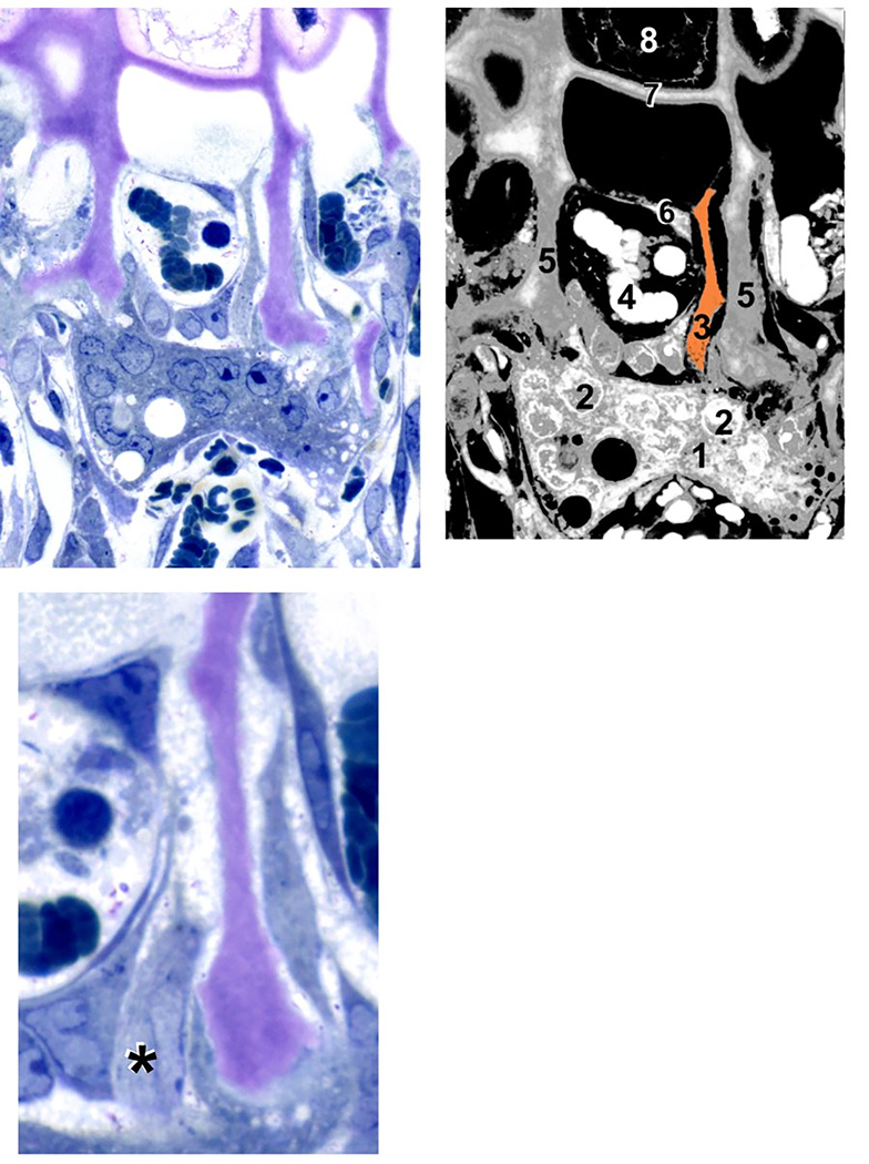
Figure 2. Septoclast in the context of the chondroosseous junction.
(A) Light micrograph of a 4-week-old wild-type rat proximal tibia, demineralized, stained with toluidine blue, 1 µm Epon section. (B) Diagram of (A) with various cellular and structural constituents numbered: 1, osteo/chondroclast; 2, some of the multiple nuclei in that cell; 3, septoclast cell body and cytoplasmic extension, pseudo-colored light orange; 4, red blood cells in budding capillary; 5, longitudinal septa of the hypertrophic cartilage zone (cartilage matrix is stained purple due to toluidine blue metachromasia); 6, endothelial cell of the capillary; 7, terminal growth plate transverse septum; 8, a hypertrophic chondrocyte in the terminal chondrocyte lacuna. (C) Serial section to (A) at higher magnification in which the septoclast cell body and nucleus are visible, but the cytoplasmic extension is out of the plane. Asterisk (*) is adjacent to the nucleus. Note that the septoclast and its capillary precede the front of osteoclasts/chondroclasts into the growth plate. Scale bar: 25 µm (A and B); 13 µm (C) (reprinted with permission from Gartland et al., 2009).

