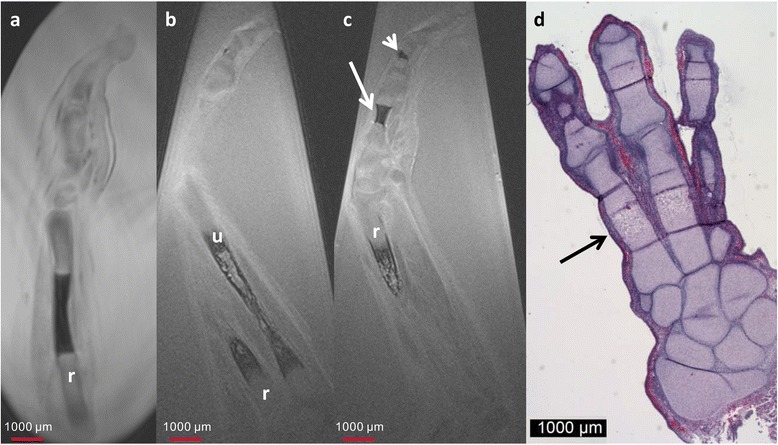
Fig. 4.

11-week GA specimen. Sagittal T2w image of a 9-week GA specimen (a), and sagittal (b) and coronal (c) T2w image of a 11 week GA specimen demonstrate growth of the ossification centers of radius (r) and ulna (o) with improved differentiation between future cancellous and cortical bone. At 11 week GA ossification of the metacarpal (arrow in c) and phalangeal bones (short arrow in c) is visible. d Corresponding HE stain with excellent correlation of the size and the location of the ossification centers (black arrow)
