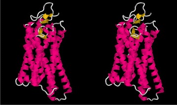
Fig. 3.

Rhodopsin structure. The eukaryotic rhodopsin structure (PDB:1gzmA) is shown as a stereo backbone cartoon as rendered by the program RASMOL [31] with secondary structures coloured as: magenta -helix and yellow -strands. The membrane runs horizontally through the middle of the molecule at right angles to the page. The seven transmembrane helices run back and forth through the membrane
