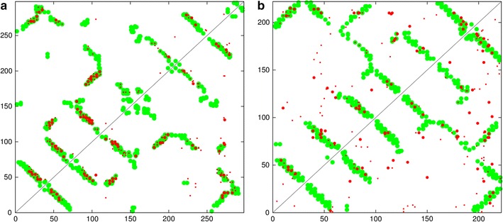
Fig. 4.

Rhodopsin contact maps. The predicted contacts (red) are compared with the observed (green) from the structure of rhodopsin (PDB code:1gzm). Top left are the top scoring 180 contacts predicted by the GREMLIN method and bottom-right, from the PSICOV program. A similar plot for the bacteriorhodopsin protein (PDB code: 2brd). The top 50 highest scoring contact predictions have a slightly larger dot and both sets omit sequence separations under five
