
Fig. 7.
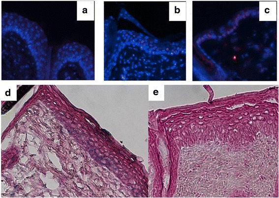
Interleukin (IL)-33 expression and promoter activation in the skin during imiquimod (IMQ)-induced skin inflammation. Immunofluorescence staining for IL-33 in skin samples 11 days after daily IMQ or Vaseline application. IMQ-treated IL-33-/- mice showed no expression of IL-33 (a), whereas IMQ-treated wild-type (WT) mice showed IL-33 expression (red) in keratinocyte nuclei (b) as did WT mice without IMQ treatment (c). On the bottom panel, the activity of the endogenous IL-33 promoter, by X-Gal staining, showed IMQ-treated IL-33-/- mice with expression in basal layers of the epidermis (d) and no detection in WT mice (e) (×200)