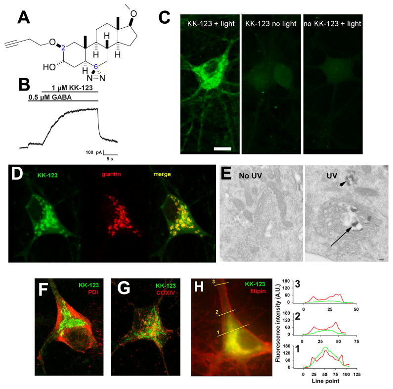
Figure 1.
Golgi-selective labeling of cultured hippocampal neurons by a bi-functional, GABA-active neurosteroid analogue. A. Structure of KK-123, showing the alkyne tag (dashed circle) for click chemistry at carbon 2 and the diazirine (dashed rectangle) at carbon 6 for photolabeling. B. Potentiation of a GABA activated current in a hippocampal neuron. The neuron was recorded in whole-cell, patch-clamp configuration, filled with a cesium methanesulfonate solution, and clamped at 0 mV. Application of 1 μM KK-123 increased the outward current in response to GABA application. C. A hippocampal culture was incubated in 1 μM KK-123 and either exposed to 365 nM UV illumination for 15 min or incubated in the dark, fixed, and processed for click cyto-fluorescence using azide-conjugated AlexaFluor 488. The panels are labeled with the experimental conditions. Only the UV illuminated condition (light) with KK-123 showed significant labeling. D. Combined click cyto-fluorescence with immunofluorescence for giantin, a Golgi-specific protein, revealed extensive co-labeling. E. Click reaction performed with azide-conjugated biotin, and subsequent processing for electron microscopy (see Methods) revealed reaction product associated with Golgi (arrow) and Golgi-related structures (arrowhead). F–H. Little co-localization with the endoplasmic reticulum marker PDI, the mitochondria marker COXIV, or the fluorescent marker of free membrane cholesterol, filipin. The right panel in H shows line scans of the red (filipin) and green (KK-123) channels to demonstrate partial overlap in labeling, but with KK-123 notably weaker than filipin in plasma-membrane areas, most notable in neurites (line scan 3).

