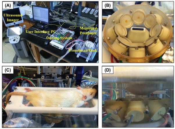
Figure 1. Histotripsy in vivo rat liver ablation experimental setup.
(A) A 1 MHz histotripsy therapy transducer with coaxially aligned ultrasound imaging probe was attached to a motorized 3D positioning system controlled using a PC console. (B) The histotripsy transducer consisted of 8 transducer elements in a ring with the imaging probe inserted in the center. (C) For treatment, an anesthetized rat was placed on a stage above the histotripsy transducer. (D) The transducer was placed inside a tank of degassed water beneath the anesthetized rat, and histotripsy was noninvasively applied to the liver through the intact abdomen.

