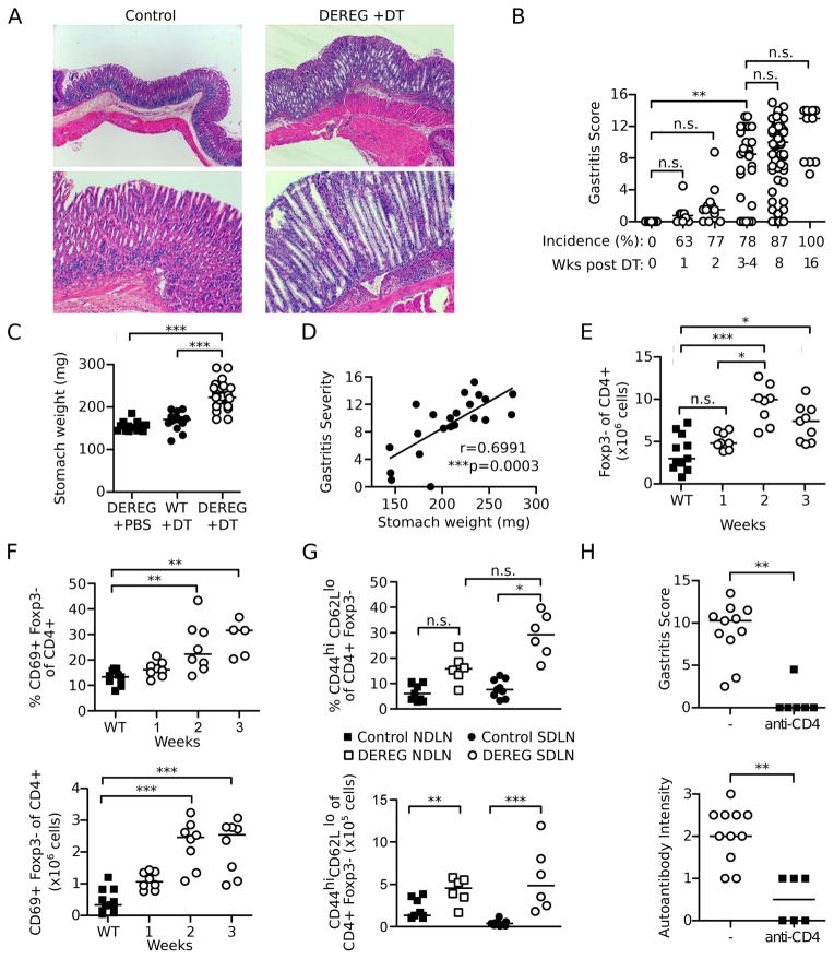
Figure 1. Transient Treg cell depletion results in AIG dependent on CD4+ T cells.
(A) Representative stomach histology showing well-organized mucosa (control mice, left) or hypertrophy, abundant mucin-positive cells, and reduction in parietal cells (Treg cell-depleted DEREG mice, right) (H&E stain; top, x40; bottom, x100). (B) Kinetics of gastritis incidences and severity after Treg cell depletion. (C) Stomach weights at 3 weeks. (D) Correlation of gastritis score and stomach weight at 3 weeks. (E) Kinetics of absolute numbers of Foxp3− of CD4+ T cells and (F) percentages (top) and absolute numbers (bottom) of CD69+ Foxp3− of CD4+ T cells in pooled lymph nodes after Treg cell depletion. (G) Percentages (top) and absolute numbers (bottom) of CD44hiCD62Llo of CD4+Foxp3− cells in the NDLN and SDLN at 3 weeks in control and Treg cell-depleted DEREG mice. (H) Gastritis score and gastric autoantibody intensity of mice at 8 weeks after Treg cell depletion with or without anti-CD4 depleting antibody treatment. Data were pooled from 2–11 independent experiments per time point. Each symbol represents an individual mouse. *p<0.05, **p<0.01, ***p<0.001; Kruskal-Wallis tests with Dunns posttests (B, C, E, F, G top); Mann-Whitney t test (G bottom, H); Spearman Correlation test (D).

