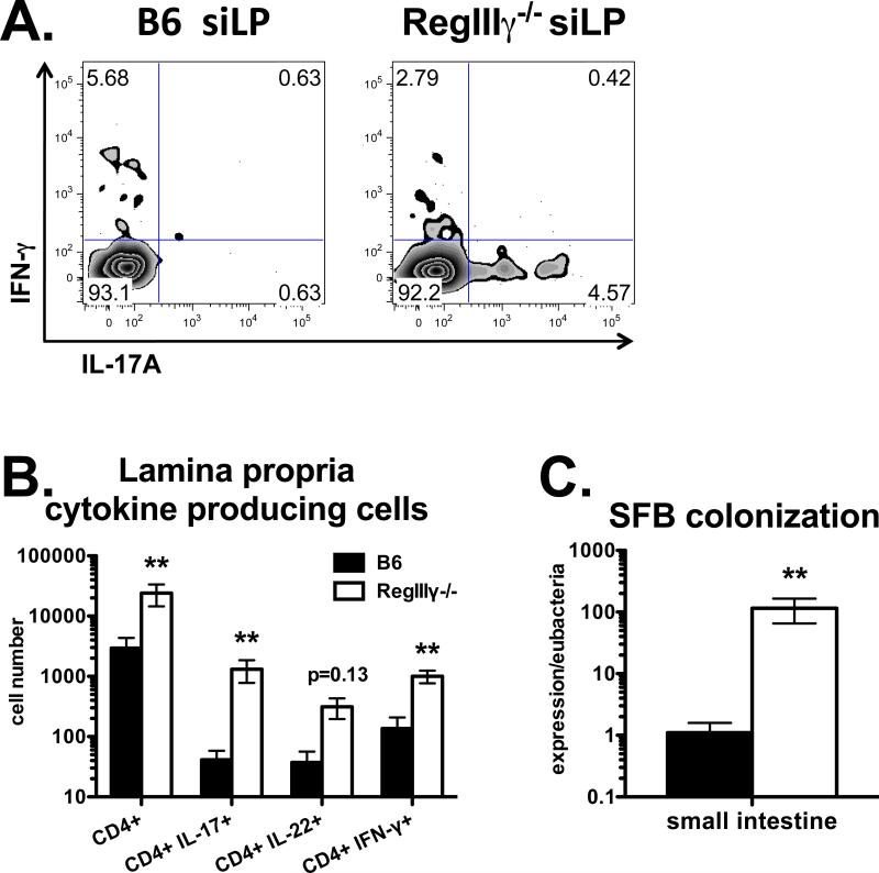
Figure 3. RegIIIγ suppresses intestinal Th17 cell priming.
Mice were injected i.p. with OVA plus LPS. Three weeks later, siLP lymphocytes were stimulated with PMA plus ionomycin. A. Flow cytometry plots of IL-17 vs. IFN-γ staining, gated on CD4 T cells. B. Total numbers of siLP CD4 T cells producing IL-17, IL-22 or IFN-γ. Data in A and B are combined from two experiments with n=6. C. SFB colonization determined by PCR from small intestinal cDNA, normalized to Eubacteria. Data in C are combined from 4 experiments with n=8-9.

