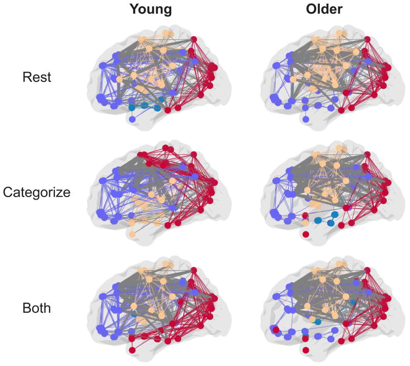
Figure 3.
Sagittal views of group partitions during resting-state (top) and the least and most demanding task conditions (middle and bottom, respectively) for young and older adults. For visualization purposes, group consensus partitions were created by averaging the AAL atlas correlation matrices across subjects in each group and thresholding at 20% of possible network connections (although note that all analyses presented in the Results were done at the individual subject level at multiple connection thresholds as described in the Materials and Methods). Within-module edges are colored to match that of nodes in their own module and between-module edges are colored gray, with lateral frontal between-module connections bolded. Older adults show more between-module lateral frontal connections at lower levels of demand (CATEGORIZE) compared to young adults. Young and older adults have similar amounts of lateral frontal connections during resting-state and the more demanding task condition (BOTH).

