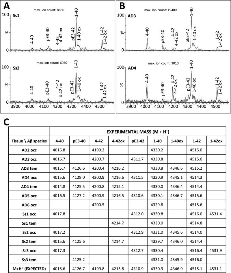
Figure 4. MALDI-TOF mass spectrometry analysis of insoluble Aβ peptides in AD and aged squirrel monkey neocortices.
Panel A: Aβ spectra obtained from occipital cortical extracts of two aged-squirrel monkey specimens, Ss1 (top) and Ss2 (bottom). Panel B: Aβ spectra obtained from temporal and occipital extracts of two AD patients (AD3, top; AD4, bottom). Labeled peaks correspond to full-length, truncated, or post-translationally-modified Aβ peptides. Panel C: Comparative list of theoretical and experimental m/z values for all AD and monkey samples tested As a whole, the spectrometric pattern of Aβ peptides in aged squirrel monkeys was similar to that of the AD group.

