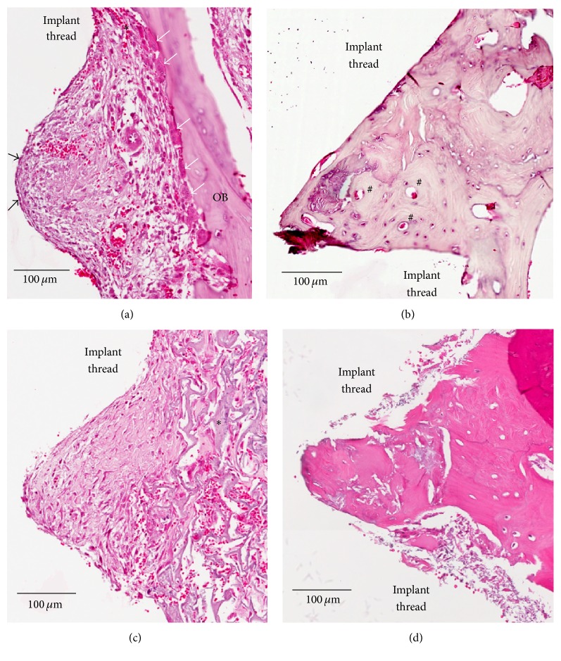
Figure 8.
Ground sections of the peri-implant tissue of GDP-fib and Ar-GDP implant surfaces after 2 and 8 weeks of healing. (a) Wound chamber of the GDP-fib implant at 2 weeks. Osteoclasts (white arrows) and osteoblasts (black arrows) were surrounded by provisional matrix, decalcified section, original mag. ×200. (b) Wound chamber of the GDP-fib implant at 8 weeks. Osteon (#) could be clearly identified, decalcified section, original mag. ×200. (c) Wound chamber of the Ar-GDP implant at 2 weeks. Woven bone (∗) formation extending into provisional connective tissue matrix was seen, decalcified section, original mag. ×200. (d) Wound chamber of the Ar-GDP implant at 8 weeks, decalcified section, original mag. ×200.

