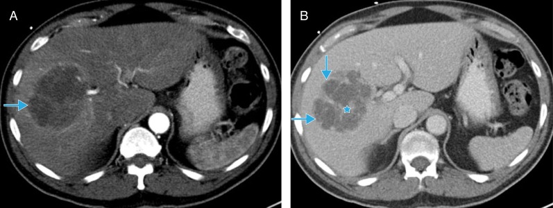
Fig. 4. Dynamic contrast-enhanced computed tomography (CT).
A. Late arterial phase CT demonstrates hypervascular, peripheral enhancement of the abscess seen in Figure 4 (blue arrow). B. Portal venous phase CT demonstrates conspicuity of internally enhancing septations (blue star), likely representing intervening hepatic parenchyma. Note the multilocular nature of the abscess, which has implications for potential treatments (blue arrows).

