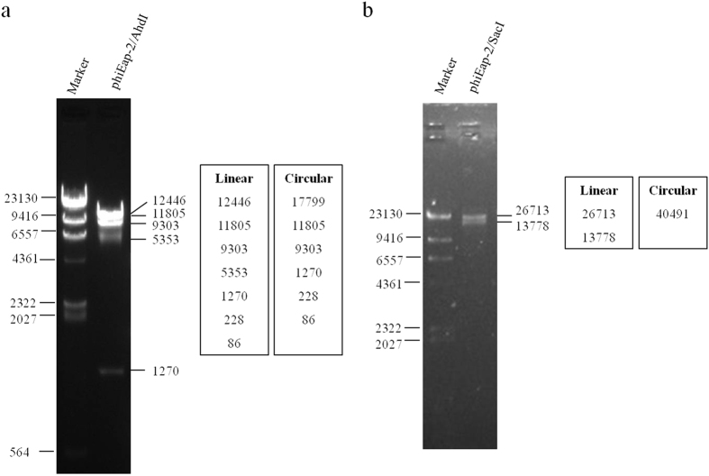
Figure 2. Restriction fragment length polymorphism analysis of phiEap-2 DNA.
Genomic DNA from phage phiEap-2 was digested with the enzymes indicated (AhdI and SacI) and run on an agarose gel (0.7%). The length of fragments generated by digestion of the linear genome or the circular genome was showed on the right side of the electrophoresis map.

