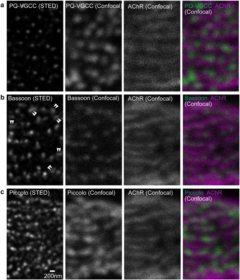
Figure 1. STED microscopy revealed distribution patterns of active zone proteins in an en face view of adult mouse NMJs.
Fluorescent immunohistochemistry was used to visualize (a) P/Q-type voltage-gated calcium channels pore forming α subunit (PQ-VGCC), and active zone proteins (b) Bassoon and (c) Piccolo in NMJs of eight-month-old wild-type mice. STED microscopy images (far left column, deconvolved images) demonstrated significantly improved resolution of these three proteins compared to confocal microscopy images (second column from the left) in single optical plane images parallel to the presynaptic membrane. Double arrowheads in the Bassoon STED image indicate examples of doublet Bassoon puncta that were not resolved in confocal images. Confocal images revealed an alignment of these proteins with postsynaptic junctional folds, which was visualized as bright lines of α-bungarotoxin labeled acetylcholine receptors (most right column, magenta = AChR, green = active zone proteins). Scale bar: 200 nm.

