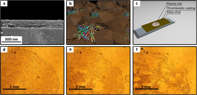
Figure 2.
SEM cross-section image of a thrombolytic coating with 12.5 wt% entrapped uPA (a); schematic representation of a magnetic thrombolytic composite (MTC) (b); scheme for thrombolytic analysis of uPA@ferria films carried out using an optical microscope (c); visualization of the plasma clot lysis process provided by MTC using an optical microscope at 0, 45, and 90 minutes, respectively (d–f), lens X10.

