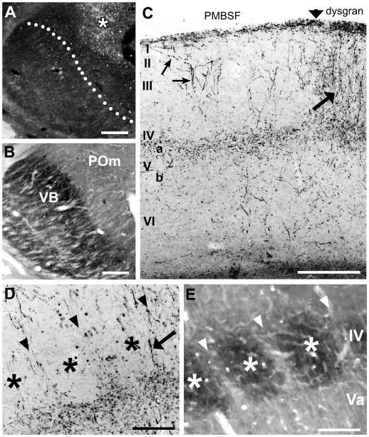
Fig. 1.
Laminar pattern of POm TCA innervation of adult mouse barrel cortex.
A, B: Photomicrographs of adjacent coronal sections through somatosensory thalamus showing a representative injection of PHA-L (asterisk) in the POm nucleus (A). The image in (A) was taken in darkfield optics, so the injection site appears white against a dark background. The adjacent section (B) was processed histochemically for cytochrome oxidase activity to indicate nuclear borders. The dotted line in (A) corresponds to the border between VPM and POm nuclei shown in (B). C, D: Photomicrographs of coronal sections through adult mouse barrel cortex showing laminar innervation patterns of PHA-L-labeled POm TCAs and terminals (dark lines and stipple). C: Pia-to-white matter image through the border (vertical arrow) between the PMBSF and the dysgranular cortex (dysgran). In the PMBSF, labeled axons formed a dense terminal plexus in layer Va; innervation of layer IV was sparse. Some axons extended superficially, and branched obliquely to enter layer I (small arrows, left). Granular and supragranular layers of the dysgranular cortex were densely innervated (arrow, right). In this and subsequent figures, Roman numbers refer to cortical layers. D, E: higher power adjacent coronal sections through layer IV showing PHA-L-labeled axons and terminals (D) or cytochrome oxidase staining (E) to indicate position of intensely reactive barrels (asterisks) and more lightly stained, intervening septa (arrowheads). Septa are narrow in mice, and few axon terminals or fibers (arrow, D) were present in layer IV. Scale bars = 250 μm in A–C; 100 μm in D, E.

