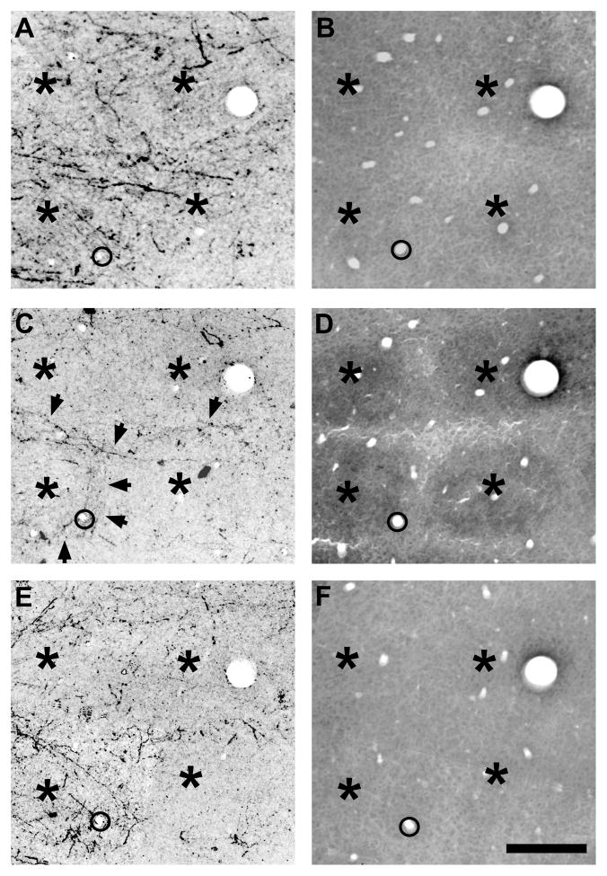
Fig. 12.
Septa-related columns of POm TCA terminations spanning layers III through Va in rats. Photomicrographs of serially-ordered tangential sections spanning lower layer III (A, B), through layer IV (C, D) to layer Va (E, F) showing PHA-L-labeled POm TCAs (A, C, E) or cytochrome oxidase staining of the adjacent sections (B, D, F) to indicate the more intensely reactive barrels in layer IV (asterisks, D). PHA-L was injected into the POm nucleus on P6, the animal was killed on P8. Asterisks indicate the barrels in layer IV (C, D), or their corresponding positions in layer III (A, B) and layer Va (E, F). In layer IV, TCAs (arrows) are largely confined to septa. Labeled TCAs remain concentrated within septal-like columns in layer III and in layer Va, with fewer axons present above and below the position of the barrels (asterisks). The circle in the lower left corner of each image shows a blood vessel common to all sections. Scale bar = 200 μm in F (applies to A–F).

