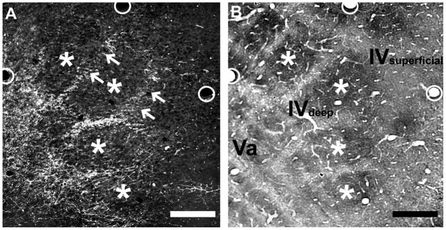
Fig. 2.
Septal-related patterning of POm TCAs in adult mouse PMBSF.
Photomicrographs of adjacent sections cut in an oblique, tangential plane through middle layers of the PMBSF, showing PHA-L-labeled POm TCAs in darkfield optics (A), or cytochrome oxidase activity (B) which reveals borders between barrels (asterisks) and intervening septa. The plane of the oblique section extends from the upper half of layer IV (layer IV superficial) through the deeper half of layer IV (layer IV deep) into layer Va. PHA-L-labeled POm TCAs formed a dense plexus in layer Va that extended into the septa at the layer IV/Va border and into the deeper half of layer IV, but the density of fibers within the septa decreased significantly in passing into the superficial half of layer IV (arrows). Very few labeled axons were present within the barrels. The circles in the two images show matched positions of the same blood vessels. Scale bars = 100 μm in A, B.

