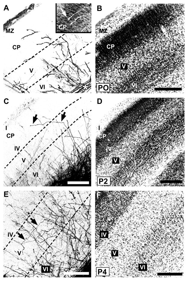
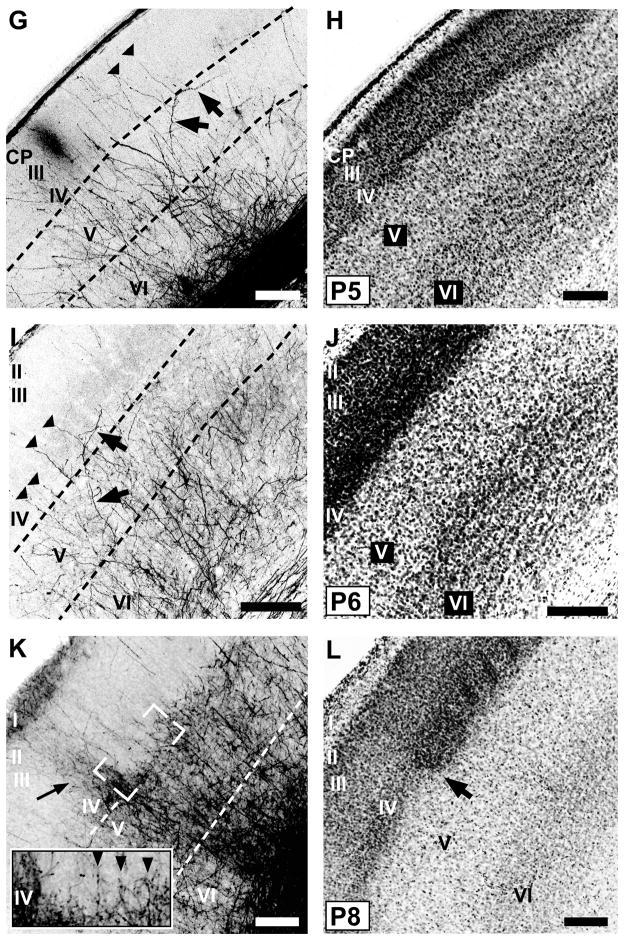
Fig. 3.
Developmental progression of laminar innervation by POm TCAs in mouse barrel cortex.
Pairs of confocal microscope images of coronal sections through developing mouse PMBSF at various postnatal ages from birth (P0) through P8 showing POm TCAs labeled by DiI (A,E,G,I,K) or DiD (C), and laminar cytoarchitecture revealed by Nissl counterstaining (B,D,F,H,J,L) of the corresponding dye-labeled sections. Dotted lines in the DiI/D images indicate borders between layers. A, B: At P0, many POm TCAs turned into barrel cortex from a subjacent stratum of labeled axons in the white matter (asterisk). Many axons penetrated the dense cortical plate (CP), and some were tipped by growth cones (inset, arrow; DiI-labeled axons are shown in white against the Nissl counterstaining). MZ, marginal zone. C, D: By P2, many radially oriented POm TCAs in layer V sent an oblique, horizontal branch into layer IV (arrows, C), which has differentiated from the CP by this age (D). E, F: By P4, overall density of labeled POm TCAs had increased in deep layers; long horizontal branches were evident in layer V (arrows, E). G, H: By P5, regularly-spaced, radially oriented branches spanned layer IV and extended into superficial layers, some of which (arrowheads, G) could be traced from long horizontal or oblique branches in layer V (arrows, G). I, J: By P6, the regularly spaced, radial branches that spanned layer IV (arrowheads, I) were evident; fewer labeled axons were found superficially in comparison with younger stages. Some oblique branches arose from layer V and traversed layer IV horizontally (arrows, I). K, L: By P8, features of an adult-like pattern emerged. Brackets (K) indicate position of image through layers IV/Va shown at higher power in inset. Regularly spaced, single fibers penetrated radially through layer IV in the PMBSF (arrowheads, inset). The layer Va plexus appeared scalloped at the border with layer IV. Adjacent to the PMBSF, labeled POm TCAs innervated densely all layers of dysgranular cortex (arrow, K). The cytoarchitectural border between the PMBSF and dysgranular cortex is shown by arrow in (L). Scale bars = 200 μm in B–L.


