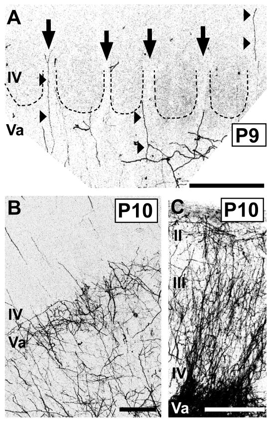
Fig. 5.
Features of the adult-like pattern of laminar innervation by POm TCAs emerge at the beginning of the second postnatal week in mice.
Confocal images of carbocyanine dye-labeled POm TCAs in middle layers of PMBSF (A, B) or in dysgranular cortex (C). A: High-power image of a section through layers IV/Va taken from a P9 mouse. Dotted lines demarcate outlines of individual barrels; the barrels are visible by refringence. Septa are indicated by arrows. A few, radially oriented DiD-labeled axons entered or traversed layer IV through the septa (arrowheads); the origins of many could be identified as extensions from oblique or horizontal branches in the layer Va plexus. In this animal, the injection was small and very few axons were labeled, affording great clarity in the pattern. B: Image taken from a P10 mouse in which the DiI injection site was larger, resulting in a greater density of DiI-labeled POm TCAs. At this stage, the dense layer Va plexus was adult-like, as was the sparse innervation of layer IV. C: Image taken from same animal as that shown in (B); the POm TCA innervation of dysgranular cortex was dense and adult-like. Scale bars = 200 μm in A–C.

