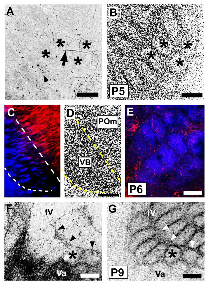
Fig. 6.
Emergence of a septal-related pattern of POm TCA innervation in developing mouse barrel cortex.
A, B: Confocal microscope images of a tangential section through layer IV taken from a P5 mouse showing DiI-labeled POm TCAs (A) and barrel/septal cytoarchitecture revealed by Nissl-counterstaining (B). Asterisks show the same barrels in the two images; barrels are also visible in (A) by refringence. Short axon-fragments were found in both barrels (arrowhead) and septa; occasional longer, horizontally oriented axons were also seen in the septa (arrow). No overt septal pattern was yet evident at this age. C, D: A representative pair of adjacent, confocal images of coronal sections through the somatosensory thalamus of a P6 mouse in which two different carbocyanine dyes (C) were used simultaneously to label VPM (DiD, blue) and POm (DiI, red) TCAs. Nuclear borders were revealed cytoarchitecturally by Nissl staining (D); dotted lines delineate the extent of the VPM nucleus. E: Confocal image of a tangential section through layer IV taken from a mouse in which VPM and POm nuclei were both labeled as shown in (C). At P6, whisker-related clusters of VPM TCAs (blue) were well-formed as expected. Sparsely-distributed POm TCAs were found in both barrels and septa. F, G: Confocal images of an oblique, tangential section spanning the upper half of layer IV (upper right in the image) through layer Va (lower left in the image) taken from a P9 mouse showing DiI-labeled POm TCAs (F) and layer IV barrel/septal cytoarchitecture revealed by Nissl counterstaining (G). By this age, the dense layer Va plexus of POm TCAs was evident, as was a mature-like septal-pattern of innervation in the lower half of layer IV, particularly along barrel rows (arrowheads). The asterisks denote the position of the same, representative barrel in the two images. Scale bars = 200 μm in A, B, D–G (that in D also applies to C).

