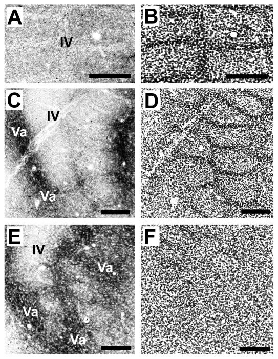
Fig. 7.
A mature-like septal-innervation pattern by POm TCAs is evident in mice by P10.
Confocal images of an adjacent series of tangential sections spanning the full depth of layer IV (A–D) through the layer IV/Va border (E,F) taken from a P10 mouse showing DiD-labeled POm TCAs (A, C, E) and barrel/septal cytoarchitecture revealed by Nissl counterstaining (B, D, F). A mature-like pattern was fully resolved by this age. Labeled POm TCAs were very sparse in the upper half of layer IV (A, B), but in passing progressively deeper, they became increasingly concentrated in the septa, particularly along barrel rows (C,D). At the layer IV/Va border, the septal-innervation pattern was prominent, with labeled POm TCAs also appearing in the position of the barrels as the plane of the section dips into the layer Va plexus (E, F). Scale bars = 200 μm in A–F.

