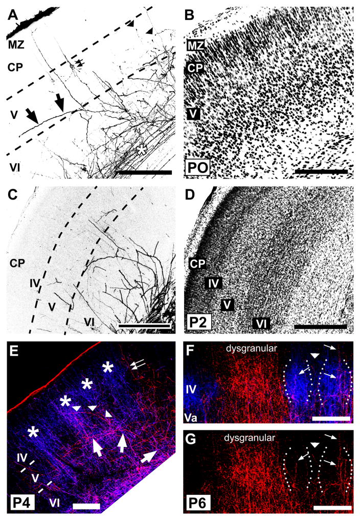
Fig. 8.
Developmental progression of laminar innervation by POm TCAs in rat barrel cortex.
Confocal microscope images of coronal sections through developing rat PMBSF at various postnatal ages showing DiD-labeled POm TCAs (A,C) and corresponding laminar cytoarchitecture revealed by Nissl counterstaining (B,D), or showing dual labeling of VPM and POm TCAs (E–G). Dashed lines in the DiD images indicate borders between layers. A, B: At P0, POm TCAs entered barrel cortex from a white matter stratum of labeled axons (asterisk). Many axons were tipped by growth cones (arrowheads), but unlike mice, few penetrated the dense cortical plate (CP). Most axons were radially oriented; some however took long, horizontal or oblique trajectories into layer V (arrows). Small double arrows indicate a retrogradely-labeled corticothalamic cell. MZ, marginal zone. C, D: By P2, labeled axons remained mostly restricted to infragranular layers and avoided layer IV, which has emerged from the CP by this age. E: By P4, whisker-related clusters of VPM TCAs (DiD, blue) within layer IV barrels (asterisks) were well formed as expected. Branches of DiI-labeled POm TCAs (red) had by this age entered and extended through layer IV, although they occupied both septa and barrels (arrowheads). Other labeled POm TCAs took long, oblique trajectories through infragranular layers, spanning several barrel-widths (large arrows). Some radial axons extended into superficial layers (small double arrows), reaching layer I. F, G: Confocal images through middle layers taken from a P6 rat in which the VPM and POm TCA projections were labeled simultaneously. In (F), the channel showing DiD-labeled POm axons (red) is merged with that showing DiI-labeled VPM TCAs (blue), while in (G), the channel showing POm TCAs is displayed separately. Labeled POm TCAs remained in both septa (arrowhead) and barrels (arrows), but they appeared to be receding somewhat from barrels in comparison with younger ages. The dysgranular cortex was densely innervated by this age. Scale bars = 200 μm (A–G).

