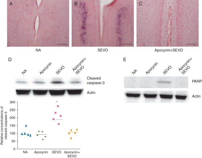
Fig 4.
Representative images showing caspase-3 activation in the retrosplenial cortex of the brain (scale bar=80 μm). Black dots indicate cleaved caspase-3-positive cells in (a) NA, (b) SEVO and (c) Apocynin+SEVO groups. (e) Representative image and relative cumulative concentrations of cleaved caspase-3 in 6 h after sevoflurane exposure (*P<0.05, NA vs SEVO, n=5 each). (e) Representative image showing expression of the apoptosis marker, PARP, in the retrosplenial cortex of the brain after sevoflurane exposure.

