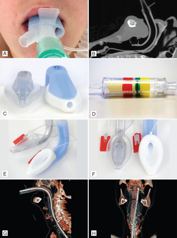
Fig 1.
(a) LMA-Protector™ in situ, showing two ports. (b) Computed tomography scan of LMA-Protector™ in situ. (c) LMA-Supreme™ and LMA-Protector™, showing the 10° slant of the tip of the distal part of the cuff. (d) Cuff Pilot Valve™. (e) and (f) LMA-Supreme™ and LMA-Protector™ lateral (e) and frontal view (f). (g) and (h) Computed tomography reconstruction of LMA-Protector™, showing tissue and gastric tube path in the lateral (g) and frontal view (h). Lateral X-ray image of LMA-Protector™ in situ.

