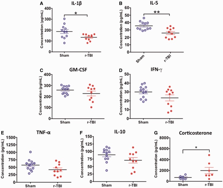
FIGURE 10.
Inflammatory cytokine profile and corticosterone levels in the plasma in an hTau mouse model of chronic repeated traumatic brain injury (r-TBI). (A, B) There was a significant decrease in IL-1ß and IL-5 observed in the plasma of injured versus sham animals. (C–F) There was a trend toward decrease in GM-CSF, IFN-γ, TNF, and IL-10 in injured versus sham animals but these did not reach statistical significance. N = 10–12 (sham/injured). (G) There was a significant increase in corticosterone in the plasma of injured versus sham animals. N = 6–8 (sham/injured); *p < 0.05; **p < 0.01.

