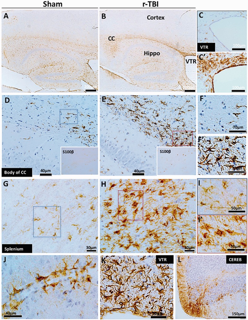
FIGURE 3.
Astrogliosis in an hTau mouse model of chronic repeated traumatic brain injury (r-TBI). (A–L). Immunohistochemical staining for GFAP (astrogliosis) demonstrates a dramatic increase along the length of the corpus callosum of injured animals compared with shams (A, B). Astroglial activation was not present in the cortex of sham or injured animals (A, B). White matter regions surrounding the ventricles also showed increased GFAP staining in the injured (C′) compared with sham animals (C). There are hypertrophic astrocytes in the body (D, E) and splenium (G, H) of the corpus callosum of injured compared with sham animals. A parallel increase in astroglial marker S100ß was also observed in the cell body of astroglial cells in the corpus callosum of injured compared with sham animals (insets of D and E). Hypertrophic astrocytes demonstrated large cell body and prominent thick processes in these regions (F vs F′, I vs I′). Prominent and hypertrophic perivascular astrocyte endfeet (strongly stained with GFAP) are seen along the length of small blood vessels in injured animals (J). High-power micrograph of periventricular region is shown in (K). GFAP staining was increased in the cerebellar molecular cell layer in injured animals (L). CC, corpus callosum; VTR, ventricles; CEREB, cerebellum; Hippo, hippocampus.

