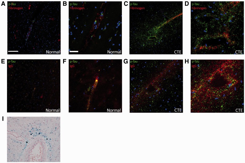
FIGURE 4.
Fibrinogen and IgG extravasation in normal brain (A, B, E, F) and in the brain of a patient with chronic traumatic encephalopathy (CTE) (C, D, G–I). (A–D) Fibrinogen (red) and p-Tau (green) expression in normal human brain (A, B) and CTE (C, D) samples. (E–H) Human IgG (red) and p-Tau (green) staining in normal human brain (E, F) and CTE brain (G, H) samples. (I) White matter perivascular hemosiderin deposition in the CTE patient brain demonstrated with Perl’s stain. (A, E, G, I) 10X objective; scale bar: 100 µm; (B, D, F, H) 40X objective, scale bar: 20 µm.

