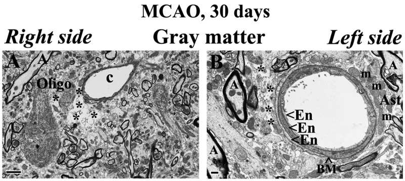
FIGURE 4.
Electron microscope examination of potential repair of microvasculature in the rat cervical spinal cord gray matter at 30 days after tMCAO. (A) Activated-appearing oligodendrocytes characterized by dark condensed cytoplasm are seen near an edematous space and damaged capillary on the right side of cord in gray matter. (B) A capillary with 3 layers of ECs and astrocyte end-feet with normal mitochondria is seen on the left cord side of gray matter. Abbreviations: En, endothelial cell; BM, basement membrane; Ast, astrocyte; m, mitochondrion; c, capillary; A, axon; Oligo, oligodendrocyte. Asterisks in A and B indicate extracellular edema. Scale bars: A = 2 μm; B = 500 nm.

