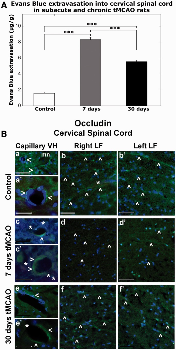
FIGURE 5.
Quantitative analysis of Evans blue (EB) parenchymal extravasation and occludin microvascular immunostaining patterns in the rat cervical spinal cord at 7 days and 30 days after tMCAO. (A) Quantitative measurement of parenchymal EB content showed higher extravasated EB levels at 7 days and 30 days post-tMCAO versus control (p < 0.001). Levels at 7 days were greater than at 30 days (***p < 0.001). (B) Immunofluorescence staining for occludin (a, a′) within ventral horn capillaries (green, arrowheads) and (b, b′) in lateral funiculus of both sides of the cervical spinal cord (green, arrowheads) show typical tight junction expression patterns in control rats. Capillaries with strong occludin immunostaining were noted near motor neurons (mn) in ventral horn (a). The EB is within microvessels in white matter lateral funiculus (b, light red, asterisk). At 7 days post-tMCAO, weak occludin immunoreactivity was observed in (c, c′, green, arrowheads) portions of numerous ventral horn capillaries with detection of perivascular EB leakage (light red, asterisks). Occludin immunostaining was identified in only a few microvessels in the lateral funiculus of both sides of the spinal cord in these animals (d, d′, green, arrowheads). At 30 days after tMCAO, limited occludin immunoexpression (e, e′, green, arrowheads) was determined in ventral horn capillaries, which some of them are leaky for EB (light red, asterisks), similar to 7 days post-stroke. Microvessels with enhanced occludin immunostaining in both sides of lateral funiculus areas were noted in these animals similar to controls (f, f″, green, arrowheads). Scale bars: a, b, b′, c, d, d′, e, f, f′ = 50 µm; a′, c′, e′ = 25 µm.

