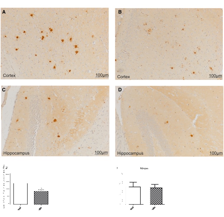
FIGURE 3.
Effect of forskolin on Aβ deposition. Representative photomicrographs of coronal sections through cortex and hippocampus show reduction of Aβ deposition following forskolin treatment. (A–D) There were numerous, relatively large Aβ plaques in the cortex of a control mouse (A) as compared with the forskolin-treated group (B). There were larger Aβ deposits in the hippocampus of a mouse from the control group (C), as compared with the forskolin-treated group (D). (E–H) Arithmetic means of plaque counts and of IR area percentages. In the cortex of mice treated with forskolin there were fewer Aβ plaques (E) and smaller IR area percentages of Aβ staining (F) than in control mice. The percentage area of Aβ in the hippocampus (H) was also reduced for the forskolin-treated group. *p < 0.05.

