
Fig. 5. Comparison of Dlx5−/− and Dlx6−/− head skeletal phenotypes and branchial arch gene expression changes.
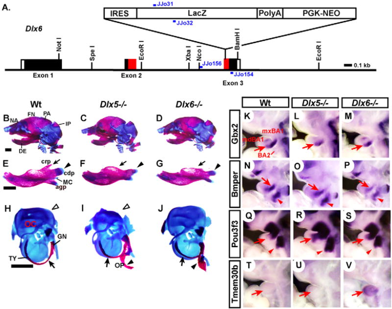
(A) Structure of the Dlx6-lacZ (Dlx6−) allele. Black boxes, exon coding region; white boxes, untranslated region; red boxes, homeodomain; blue bars, PCR primers for genotyping. (B–J) Head skeleton and skeletal elements of E18.5-P0 mice stained with Alcian Blue (cartilage) and Alizarin Red (bone). (B–D) Lateral views of the whole head. (E–G) Dentaries. (H–J) Otic capsules and associated skeletal elements. Arrows and arrowheads indicate the skeletal abnormalities of the mutants; see text for details. (K–V) Lateral views of E10.5 mouse embryos processed by whole-mount in situ hybridization. Arrows and arrowheads, downregulation (K–P) or upregulation (Q–V) of expression in the mutant mdBA1 and BA2, respectively. agp, angular process; cdp, condylar process; crp, coronoid process; GN, gonial; IP, interparietal; MC, Meckel’s cartilage; NA, nasal bone; OC, otic capsule; OP, os paradoxicum; TY, ectotympanic; for remainder, see legend to Fig. 4. Scale bar: 1 mm.