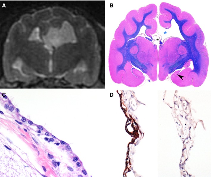
Figure 4.

Brain of 1 affected cat illustrating 2 communicating cysts. Coronal T2W magnetic resonance image (A), subgross image of the corresponding transversal section stained with luxol fast blue/hematoxylin and eosin (B), hematoxylin and eosin (HE) of cyst lining (C), and glial fibrillary acidic protein (GFAP; left) and pancytokeratin (Lu5; right) immunohistochemistries (D). The first cyst is a distended thin membrane that communicates with the lateral ventricle and is bordered by the cerebral cortex and white matter consistent with fimbria (B, blue asterisk), and causes a midline shift noted on imaging. The second cyst is a dilation of the suprapineal recess and communicates with the third ventricle (B, black smaller asterisk). The hippocampus in affected cats is often hypoplastic and displaced caudally. As noted in this animal, the septal hippocampus is absent and there is severe hypoplasia of the temporal hippocampus (B, black arrowhead) in the affected hemisphere (right side). Also note that the cingulate is rotated medially (just above the blue asterisk). Also notable is the lack of corpus callosum (interrupted by the enlarged pineal recess, black asterisk) and interthalamic adhesion. The cyst is linked by membrane of variably ciliated polygonal cells (C) that are GFAP positive and cytokeratin negative, and rest upon a GFAP negative fibrovascular membrane consistent with an ependymal cyst (D). (C) HE, ×600. (D) Immunohistochemistry for GFAP [left] and pancytokeratin Lu5 [right], ×400.
