
Figure 2.
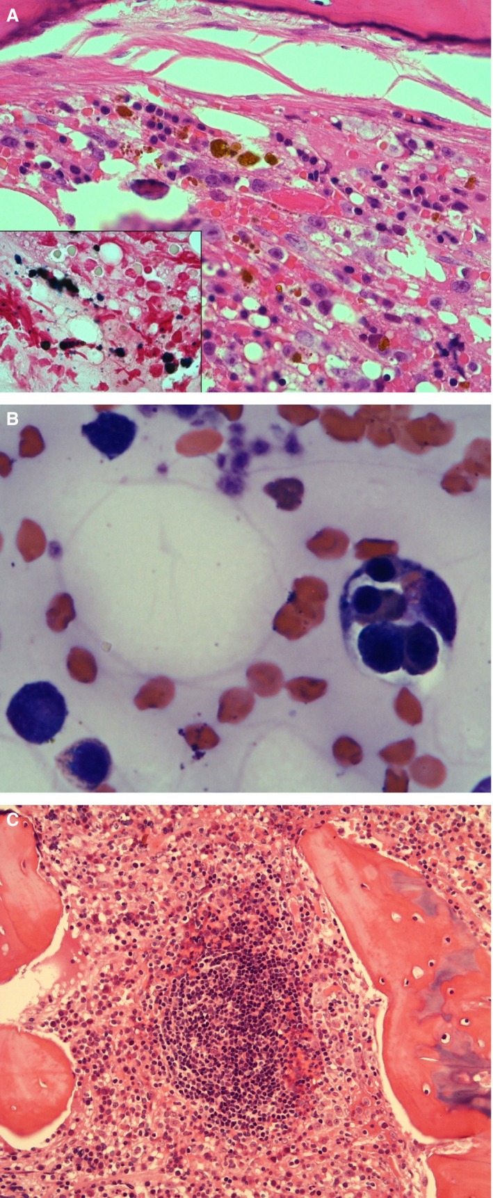
(A) Bone marrow core biopsy section of a cat with numerous foci of iron (H&E stain, 400x). Inset: stainable iron in the same sample (Perl's Prussian Blue stain, 1000x). (B) Bone marrow aspirate from a cat. Macrophage on the right with four intact erythroid precursors phagocytosed, including a basophilic rubricyte, a polychromatophilic rubricyte, and two metarubricytes (Modified Wright's stain, 1000x). (C) Prominent lymphoid follicle in a core biopsy section of feline bone marrow (H&E stain, 200x).