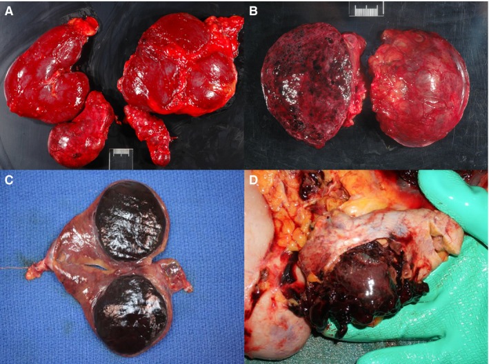
Figure 1.

(A) Left and right kidney and adrenal gland of a horse showing pheochromocytoma of the right adrenal gland. (B) Mottled, cut section of the pheochromocytoma seen in image A. (C) Depicted is a well‐demarcated pheochromocytoma with associated compression and atrophy of the adjacent adrenocortical tissue on cut section of an adrenal gland from an adult horse (photo credit Dr. Suzanne Stewart). (D) Ruptured pheochromocytoma causing hemoperitoneum in adult horse (photo credit Dr. Suzanne Stewart).
