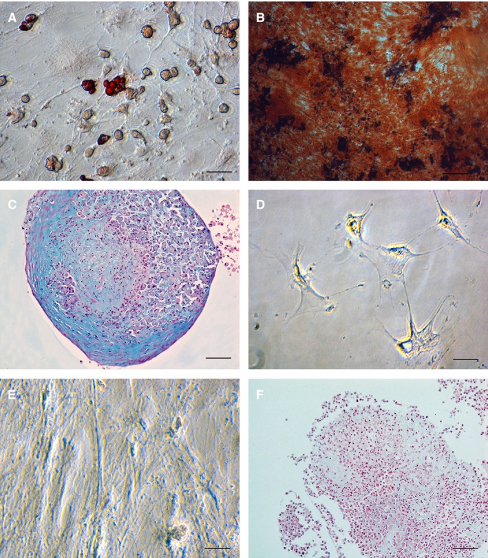
Figure 1.

Representative photomicrographs illustrating differentiation characterization of a mesenchymal stem cells cell line isolated from adipose tissue obtained from a 2 year old, female mixed breed cat (magnification 40×). (A) Lipid droplets in differentiated cells are stained red with Oil‐o‐red demonstrating adipogenesis, (B) Red‐colored calcium deposits inside cells stained with Alizarin red stain demonstrating osteogenesis, (C) Blue‐colored in glycosaminoglycan deposits in cells stained with Alcian Blue stain after chondrogenic differentiation. (D–F) Control photomicrographs after incubation of the same cell line in KNAC medium and stained with Oil‐o‐red (D), Alizarin red (E) and Alcian Blue (F). Note lack of uptake of the stains in both micrographs (D,E) and no blue staining extracellular matrix in the chondrogenic control stain (F).
