
Figure 2.
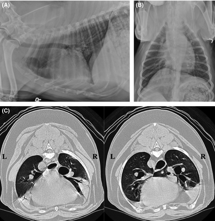
(A and B) Right lateral and dorsoventral radiographs from a dog subsequently diagnosed with diffuse bronchiectasis based on CT and bronchoscopy. Thoracic radiographs reveal mild bronchial wall mineralization, heterotopic bone formation, and mild interstitial pulmonary densities compatible with normal age‐related changes. Bronchiectasis is not identified. (C) Computed tomographic images from the dog in 2A and B. Heavy alveolar infiltrates are present in the ventral aspects of the right cranial, and right middle lung lobes, as well as the caudal subsegment of the left cranial lung lobe. In these images, bronchi to the right cranial and right middle lobes are moderately dilated, and do not taper appropriately.