Figure 4.

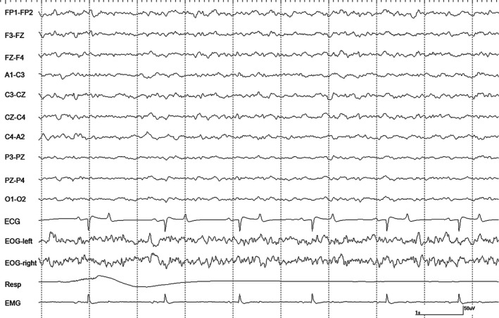
An epoch of EEG from horse #1 during halothane anesthesia at 1.2 MAC during controlled ventilation. Gain calibration is shown for EEG and EOG tracings only, others vary.

An epoch of EEG from horse #1 during halothane anesthesia at 1.2 MAC during controlled ventilation. Gain calibration is shown for EEG and EOG tracings only, others vary.