
Figure 4.
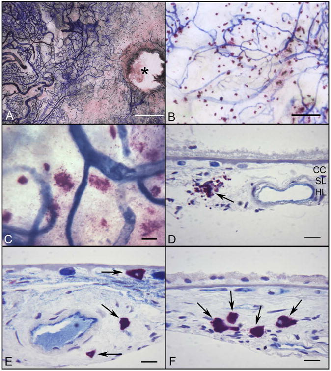
Flat mount preparation of APase/NSE incubated choroid from a subject with GA (Subject#30) (A) There is a large area of CC loss (APase−) around the optic disc (*) and in the submacular region of the choroid. (B) Many degranulated MCs are associated with areas of RPE atrophy and CC degeneration. (C) At higher magnification, most MCs are degranulated. (D-F) Histological sections from same choroid demonstrate the degranulated MCs (black arrows) aggregating in choroidal stroma in a region of RPE atrophy with a thin basal laminar deposit (D) and in the wall of an artery with atherosclerotic changes (E). MCs are increased in number but in their normal position in Sattler's and Haller's layers in a region without RPE atrophy (F). (Bar=1mm in A, 200μm in B, 50μm in C, and 20μm in D-F)