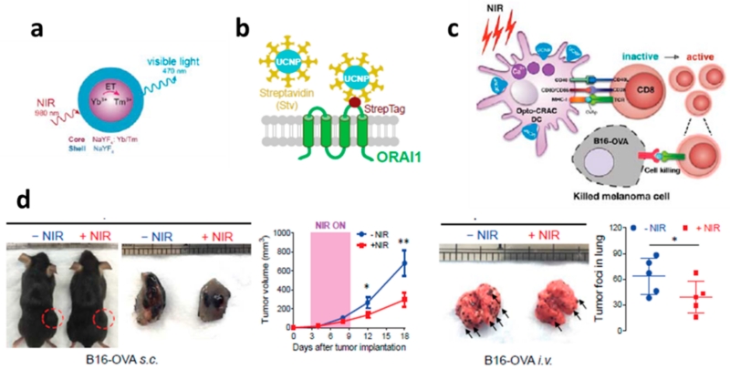
Figure 4.
(a) Schematic illustration of the core/shell structure and the energy transfer between lanthanide ions in the NaYF4:Yb,Tm/NaYF4 core/shell upconversion nanoparticles. (b) Schematic showing the interaction between streptavidin-coated upconversion nanoparticles (UCNPs-Stv) and the engineered ORAI1 Ca2+ channel that harbors a streptavidin-binding tag (StrepTag) in the second extracellular loop. (c) Scheme showing that an NIR-stimulated Ca2+ influx in Opto-CRAC DCs prompts immature DC maturation and OVA antigen cross-presentation in order to activate and to boost antitumor immune responses that are mediated by OT-1 CD8 T cells (cytotoxic T lymphocytes, CTLs). This action results in sensitizing tumor cells to OVA-specific, CTL-mediated killing in the B16-OVA melanoma model. The OVA peptide (OVAp, 257SIINFEKL264) is used here as a surrogate tumor antigen. (d) Tumor-inoculated sites (left) were isolated from tumor-bearing mice (n = 5) shielded or exposed to NIR, and tumor sizes (mm3) were measured at the indicated time points, as shown in the growth curve (right) after tumor implantation. Representative lungs with melanoma metastases (left) were isolated from tumor-bearing mice that were shielded or exposed to NIR. The histogram represents the counted numbers of visible pigmented tumor foci (denoted by the arrows) with pulmonary melanoma metastases on the surface of lungs (right panel; n = 5 mice). Reprinted with permission from ref 12. Copyright 2015 eLife Sciences Publications.

