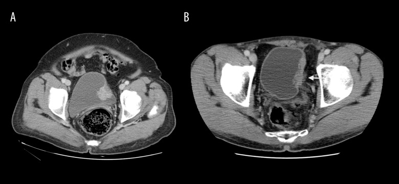
Figure 3.
Image in a 63-year-old woman with high-grade papillary urothelial carcinoma and submucosal infiltration without perivesical invasion (A). The axial 60-second delay CT image shows regular border between tumour and perivesical fatty layer. Images in a 56-year-old man with high-grade invasive papillary urothelial carcinoma with invasion of the muscle layer and perivesical fat tissue (B). On 60-second delay CT image, spread of bladder cancer to perivesical fatty tissue is seen (white arrow).

