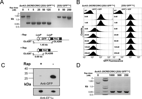
Figure 1.

Validation of inducible diCre in L. mexicana: conditional deletion of GFP in promastigotes and amastigotes.
A. Gene excision analyzed by PCR amplification. Schematic (lower) shows the SSU GFP Flox locus and the recombination event expected after treatment with rapamycin (Rap). (upper) PCR amplification with oligonucleotides 4287 and 4288 from experimental (Δcrk3::DICRE/CRK3 [SSU GFP Flox]) and control [SSU GFP Flox] promastigotes at 5 days post‐treatment with different concentrations of rapamycin.
B. Flow cytometry assessment of GFP intensity of experimental and control promastigotes incubated in the presence or absence of rapamycin for 5 days.
C. Western blotting analysis with anti‐GFP and anti‐EF1α loading control antibodies of protein extracted from experimental promastigotes grown for 5 days in the presence or absence of 100 nM rapamycin.
D. PCR analysis of GFP Flox loss (as described in A) in amastigotes after 24 h rapamycin treatment (0–1000 nM), followed by 120 h infection in bone‐marrow derived macrophages. Lane 2 contains a 1 kb+ DNA ladder.
