
Figure 3.
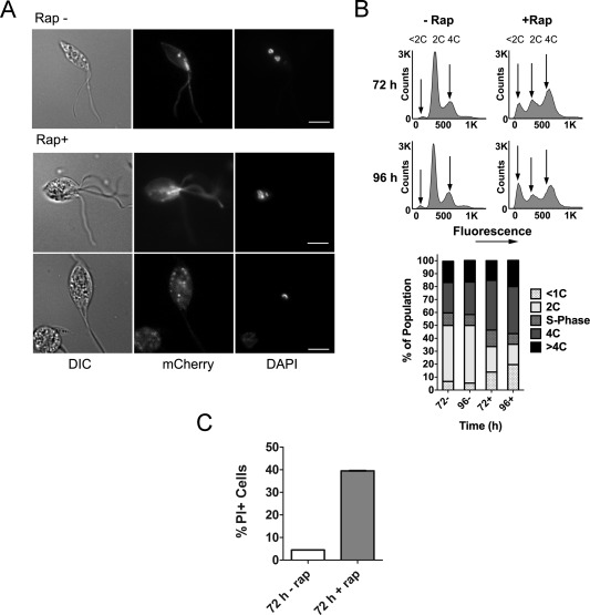
Analysis of CRK3 deficient promastigotes.
A. Representative images of cells grown in the absence (top) or presence (bottom two rows) of 100 nM rapamycin for 96 h. Promastigotes (clone 2) were stained with DAPI to observe nuclear and kinetoplast content alongside mCherry expression by fluorescence microscopy. Scale bar represents 5 μm.
B. (upper) DNA content analysis of clone 2 promastigotes at 72 and 96 h post‐treatment. Cells were fixed with methanol and stained with propidium iodide for flow cytometry analysis of 100,000 cells to examine nuclear content. Arrows indicate the positions of cells in G1 phase (2C), in G2/M (4C) and low DNA content associated with increased incidence of <1C zoids. (lower) Graphical representation of the DNA content of each population based on the flow cytometry plots.
C. The viability of cells grown in the absence (−) or presence (+) of 100 nM rapamycin for 72 h. Promastigotes (clone 2) were incubated with 5 µg mL−1 propidium iodide (PI) for 15 min and analyzed by flow cytometry. A heat lysed (HL) control in which half the sample was lysed by incubation at 70°C for 3 min was included to enable an appropriate live/dead gate to be drawn. Numbers represent the percentage of cells assessed as PI positive (PI+) based on the HL control. Data shown are the means of three technical replicates, data are representative of two independent experiments.