
Figure 3.
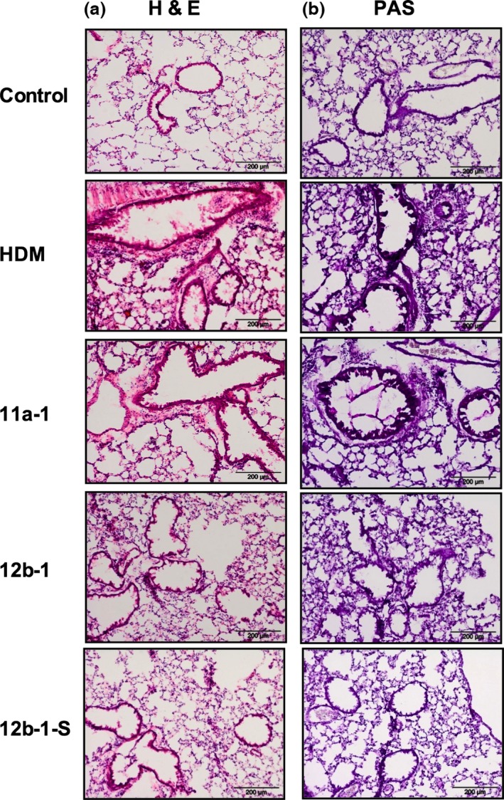
Histochemical staining of lung tissue from mice treated with SMA 11a or 12b at different time points. Alveolar regions of lung sections (7 μm) stained with H&E (a) and periodic acid‐Schiff (PAS; b) from C57BL/6 mice. Three different regions of lungs were examined in mice from two independent experiments, and representative figures are displayed. The control group was not exposed to any treatment. The HDM group represents mice exposed to HDM extracts in vehicle only. 11a‐1 and 12b‐1 represent groups of mice treated with either 11a or 12b at 1 μg per injection throughout the model. 12b‐1‐S represents a group of HDM mice treated with 12b prophylactically at the sensitization stage only. Sections were examined using an Olympus BX41 light microscope at ×40 magnification and scale bars represent 200 μm.