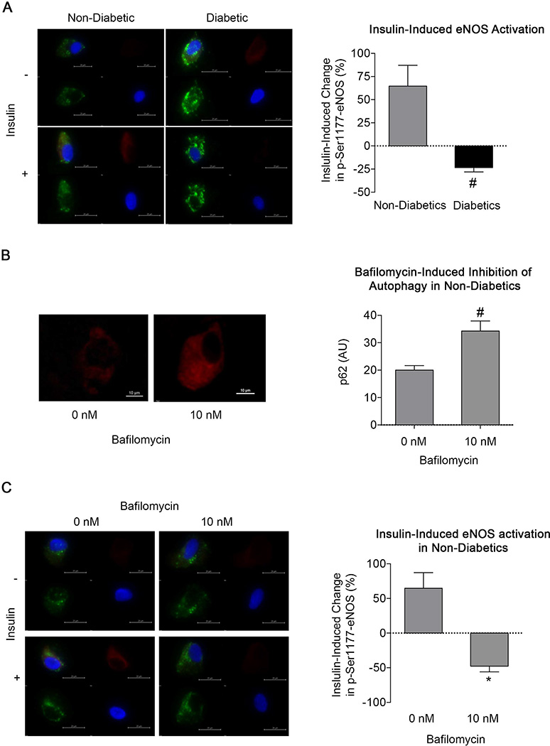
Figure 5.
Inhibition of autophagy in endothelial cells from non-diabetics impairs endothelial function. A: Insulin-induced activation of eNOS by phosphorylation at Ser1177 (red channel) was measured in endothelial cells from non-diabetics and diabetics. Also shown are vWF (green channel), DAPI (blue channel) and merged images (upper left).Cells from diabetic subjects displayed impaired activation (#P<0.01). B: Treatment of endothelial cells isolated from non-diabetic subjects (n = 6) with 10 nM bafilomycin for 60 minutes increased p62 levels, indicating impaired autophagy (#P≤0.01). C: Bafilomycin impaired insulin-induced eNOS activation (red channel) in endothelial cells from non-diabetics (*P<0.05). Also shown are vWF (green channel), DAPI (blue channel) and merged images (upper left).

